Identification guide
Lobster Mushroom Identification Guide
How to recognise Hypomyces lactifluorum in the field: a bright orange parasitic ascomycete crust that engulfs and reshapes a host Russula or Lactarius, read through its pimpled surface and white brittle interior.
What You Are Actually Looking At
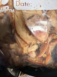
The first thing to understand is that the Lobster Mushroom is not a mushroom in the ordinary sense at all. Hypomyces lactifluorum is a parasitic ascomycete that colonises the fruitbody of another fungus, growing over it as a dense orange rind, obliterating its gills, arresting its growth and warping it into a hard, contorted shape. What you pick up is a composite object: a host fruitbody, usually a brittle-fleshed Russula or Lactarius, wearing a coat of parasite.
That means the identification has two halves. You read the orange crust to identify the parasite, and you read the white body underneath to confirm what kind of host it grew on. Neither half alone is enough.
The First Impression and the Setting
The usual setting is North American forest in summer or autumn, a week or so after sustained rain: conifer stands of hemlock, spruce and fir, and mixed conifer-hardwood woods, wherever thick-fleshed white Russula species fruit. The ground is duff, needle litter and moss.
What catches the eye is a lump of intense orange breaking the surface of the litter, often only partly emerged, with the ground heaved and cracked around it in the way that a rising Russula heaves it. The colour is startling for a forest floor: bright orange, orange-red, sometimes deep vermilion, fading to cream or whitish where the fruitbody is buried or shaded. Many specimens must be excavated rather than picked, because half the object is under the surface.
Step One: Read the Crust with a Lens
Run a thumb over the orange surface. It is not smooth. Under a hand lens it resolves into a dense pavement of minute pimples or pustules, each one a flask-shaped perithecium embedded in the parasite layer, and each with a tiny darker pore at its apex. The overall feel is of very fine sandpaper or of pebbled leather.
That pimpled texture is the single most useful field character, and it is the reason the surface has a slightly matt, granular look rather than the waxy or fibrous surface of a normal cap. Older material darkens towards orange-brown or brick, and the pustules become coarser and more crowded.
Step Two: Read the Underside
Turn the object over. On an ordinary agaric you would find a fan of thin blades. Here the gills have been overrun. What you see instead is one of three things: a nearly smooth orange surface where the gills have been completely engulfed; a set of low, blunt, irregular ridges and folds that mark where gills used to be; or, on a lightly colonised specimen, shallow parallel lines like the ghost of a gill pattern under a coat of paint.
Nothing on the underside is blade-like, nothing is free-standing and nothing could be counted. That obliteration is diagnostic of a Hypomyces infection rather than of any free-living orange fungus.
Step Three: Read the Shape, Stipe and Base
The overall form is distorted. Caps become deeply funnel-shaped, lobed, cracked and asymmetric, with torn margins, because the parasite stops normal expansion partway through. The stipe is short, thick, often eccentric or bent, and frequently swollen at its junction with the cap. The base is abrupt and usually still white or cream where soil and litter cling to it, and it is often the only part of the composite that shows the host's original colour.
The whole thing is heavy and hard for its size, denser and firmer than an uninfected agaric, and it holds together when handled roughly.
Step Four: Cut It Lengthways
A vertical section is the most informative single act in this identification. You will see a thin orange rind, roughly one to two millimetres thick, over a mass of dense white flesh. The boundary between the two is sharp, not gradual.
That white interior is host tissue, and it should tell you about the host. Press it and break it: brittle-fleshed hosts in the Russulaceae give a granular, chalky fracture rather than a fibrous tear, because their flesh contains spherical cells among the hyphae. On a fully colonised fruitbody a Lactarius host will usually not weep latex, because the parasite has arrested the host's tissue, although a partly colonised one sometimes still shows a bead or two at the cut.
Older specimens develop dull blotching within the white flesh and eventually soften from the inside while the orange rind remains firm.
Spores: Why There Is No Spore Print
A Hypomyces produces no free hymenium, so there are no gills or pores dropping spores, and a spore print in the ordinary sense cannot be taken. Do not waste an evening trying.
Instead, the spores are read directly. Scrape or slice a fragment of the orange crust, mount it in a drop of water under a coverslip, press gently, and examine it. The pressure ruptures the perithecia and releases their contents. Look for asci and, more usefully, for the ascospores: they are colourless, spindle-shaped, pointed or beaked at both ends, roughened or warted, and divided by a single central cross-wall into two cells. They are large, in the region of 30-40 microns long by 4.5-8 microns wide. Two-celled, warted, sharply pointed ascospores from an orange perithecial crust is the microscopic signature of the genus, and the size range narrows it to this species.
Substrate, Season and Ecology
The substrate here is another fungus. Hypomyces lactifluorum attacks thick-fleshed, white-fleshed members of the Russulaceae, most characteristically Russula brevipes and stout Lactarius species. It therefore appears exactly where those hosts appear: in the duff of conifer and mixed forest, often on slopes and along old trails where the host is abundant.
Infection begins early, while the host fruitbody is still a button beneath the litter, which is why the host never reaches its normal shape. The season follows the host's season, from midsummer through autumn, and depends heavily on sustained soaking rain a week or two earlier. It is chiefly a North American fungus, with the greatest abundance in western and north-eastern forests, and it recurs in the same patches over years because the host's mycorrhizal partner and the parasite persist together in the same ground.
Separating the Closest Look-alikes on Morphology
Hypomyces macrosporus is the nearest relative, growing on the same kinds of host and producing a similar crust. It tends to be duller, paler and more orange-brown, and it is separated with confidence only on ascospore dimensions, which are appreciably larger.
Hypomyces luteovirens also grows on Russula, but its crust is yellow to yellow-green, it is largely confined to the gills and underside, and it leaves the host's shape more or less recognisable.
Hypomyces chrysospermus attacks boletes rather than gilled fungi, so the host has a pore surface, and its colour sequence runs white, then powdery golden-yellow, then reddish-brown.
An uninfected Russula brevipes is the control specimen: white or dingy cream all over, with real, closely spaced, brittle gills, a short thick stipe and no orange rind whatsoever.
Orange chanterelle-like fungi including Cantharellus cinnabarinus and Turbinellus species share the colour, but they have blunt, forking, wax-like ridges or genuine folds running down the stipe, fibrous rather than chalky flesh, no perithecial pimples and no white brittle core under a rind.
Aleuria aurantia is a thin, brittle, orange cup fungus sitting on bare soil, quite unlike a heavy, distorted, rind-covered lump.
Field Checklist
A hard, heavy, distorted orange object heaved up through forest duff; bright orange to vermilion rind with a finely pimpled, sandpapery surface under a lens; gills replaced by blunt ridges or a smooth surface; short, thick, warped stipe; a sharp boundary in section between a thin orange rind and dense white brittle host flesh; two-celled, pointed, warted ascospores in place of a spore print; summer to autumn in conifer and mixed forest where thick white Russula species fruit.
Common questions
Why can I not take a spore print from a Lobster Mushroom?
How do I know a host was involved rather than this being a fungus in its own right?
Where and when should I be searching?
What is the ecological relationship at work here?
Full reference entry for Lobster Mushroom
Cap and fruiting body, spore print colour, substrate, season, ecological role and the species most often confused with it.
Open the Lobster Mushroom entry →