Identification guide
Spring Polypore Identification Guide
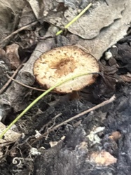
How to recognise Lentinus arcularius on fallen hardwood sticks: a small tan funnel on a central stalk, with big radially elongated decurrent pores and a fringe of stiff hairs around the cap margin.
The First Impression
Spring, a damp deciduous wood, and a fallen branch or a scatter of half-buried sticks in the leaf litter. Standing up from the wood are small tan parasols on thin tough stalks, 2 to 4 cm across, each with a dimpled or funnel-shaped centre. They look like miniature versions of a gilled mushroom until you turn one over and find, instead of gills, a coarse white honeycomb of angular pores running down onto the stalk.
That single flip — expecting gills, finding large angular decurrent pores on a small stalked mushroom — is the moment the identification really begins.
Reading the Cap
Caps are 1-5 cm across, occasionally up to 8 cm. They are circular and centrally attached, at first convex with a slight central dimple, quickly becoming depressed and finally shallowly funnel-shaped, so that the profile is a small vase on a stick.
The surface is dry and matt, yellow-brown, tawny, ochre or reddish tan, usually darker over the centre and paler at the edge, and it is covered with fine flattened scales or radiating fibrils that give a slightly roughened texture under a lens. In old, weathered specimens the colour fades to dull buff and the scaliness is worn away.
Then check the margin, which carries the best single field character. On a fresh, well-grown specimen the cap edge is fringed with a row of stiff, coarse, dark hairs standing out from the rim like eyelashes. They can be seen with the naked eye if you hold the cap against the light, and are unmistakable under a lens. These cilia wear off in old or storm-battered fruit bodies, so always examine the youngest specimens on the stick.
The Pore Surface
The pore layer is the second anchor. Pores are white to cream, becoming pale buff or slightly tan with age. They are large — around 1-2 per millimetre, individual pores easily 0.5-2 mm long — and distinctly angular, often hexagonal, and clearly elongated in a radial direction, so the surface looks like a stretched honeycomb rather than a field of round holes.
Attachment is strongly decurrent: the pore layer runs a noticeable distance down the top of the stalk before ending, and the pores nearest the stalk are the most elongated of all. The tube layer itself is very thin, usually only 1-2 mm deep, sitting directly on the thin cap flesh.
Stalk, Flesh and Texture
The stalk is central, sometimes very slightly off-centre, 1-5 cm long and only 1-4 mm thick. It is tough and pliable, pale tan to brown, often darker toward the base, and its surface is finely scurfy or minutely scaly, sometimes with a brownish felty tomentum at the very base where it grips the wood. Critically, it does not have a sharply demarcated black lower half.
Break a cap in half. The flesh is white, extremely thin, and tough and leathery rather than fleshy. It bends before it breaks, and it does not crumble. Fresh material is pliant; dried material becomes hard and horny, and rehydrates and revives after rain, so you can find the same fruit body in usable condition weeks after it appeared. There is no colour change on cutting, and the odour is faint and mushroomy.
That toughness has a microscopic explanation worth knowing: the hyphal system is dimitic, combining generative hyphae with thick-walled skeletal-binding hyphae.
Taking a Spore Print
Small polypores are fiddly to print, but it is worth doing because the result is diagnostic at the group level. Cut the stalk short, lay the cap pore-side down on a dark card or a glass slide, and cover it with a cup overnight to keep the humidity up. Because the pore surface is uneven, press it gently flat against the surface.
The print is white. Use a dark background, since a white deposit is nearly invisible on paper. Under the microscope the spores are smooth, thin-walled, hyaline, and distinctly cylindrical to sausage-shaped, roughly 7-11 x 2-3 micrometres — a shape typical of the stalked polypores.
Substrate, Season and Ecology
This is a wood-decay fungus causing a white rot, meaning it breaks down lignin as well as cellulose and leaves the decayed wood pale, soft and fibrous. It works on small-diameter dead hardwood: fallen twigs, sticks and branches lying on or half-buried in leaf litter, occasionally attached dead branches still on the tree, and sometimes larger logs. Oak, maple, beech, birch, willow, elm and many other broadleaves all serve.
The season gives the species its common name. It is one of the earliest polypores of the year, fruiting from spring into early summer, with sporadic later appearances in wet autumns. Several fruit bodies typically arise along the same stick, and the mycelium can produce successive flushes from one branch across a season.
Separating the Look-Alikes on Morphology
Lentinus brumalis, the Winter Polypore. The nearest relative and the main confusion. Its pores are small and round, about 2-4 per millimetre, and are not radially elongated; the cap is greyer brown and its margin lacks the conspicuous stiff fringe; and it fruits in the cold months from late autumn through winter into early spring.
Neofavolus alveolaris, the Hexagonal-pored Polypore. Pores are even larger and more strikingly hexagonal, but the cap is orange to reddish orange with fine scales, the fruit body is often kidney-shaped rather than circular, and the stalk is short and lateral or strongly off-centre. The cap margin is not ciliate.
Cerioporus varius. The stalk has a sharply defined black lower portion, and the pores are minute and round, giving a smooth-looking cream underside.
Picipes badius. Much larger, with a shiny chestnut to nearly black cap, a black stalk, and very fine pores.
Cerioporus squamosus, Dryad's Saddle. Also a spring fungus, but enormous by comparison, with a broad fan-shaped cap covered in large flat brown scales, a thick lateral stalk with a black base, and large angular pores.
Lentinus tigrinus. Similar stature and habitat, similar scaly cap, but the underside bears true gills with toothed edges rather than pores.
Small Coltricia species. Superficially similar stalked funnels, but they grow from soil rather than wood and have brown flesh and a brown pore surface.
Quick Field Checklist
- Spring to early summer, on dead hardwood sticks and branches in litter
- Cap 1-5 cm, circular, centrally depressed to funnel-shaped, tan with fine scales
- Margin fringed with stiff dark hairs on fresh specimens
- Pores white, large, angular to hexagonal, radially elongated and decurrent
- Stalk central, thin, tough, scurfy, without a black base
- Flesh thin, white, tough and leathery; revives when rewetted
- Spore print white; spores cylindrical
Common questions
What is the single best field character for Spring Polypore?
What does it grow on and when?
How do I separate it from the Winter Polypore?
What is its role in the woodland?
Full reference entry for Spring Polypore
Cap and fruiting body, spore print colour, substrate, season, ecological role and the species most often confused with it.
Open the Spring Polypore entry →