Identification guide
Chaga Identification Guide
How to recognise Inonotus obliquus on a living birch: the cracked black sterile conk with its rusty interior, the rarely seen poroid fertile stage under the bark, and the burls and hoof brackets that mimic it.
Standing in a Birch Wood
Chaga announces itself as a wrong-looking lump on an otherwise sound-looking tree. You are in a northern birch stand, scanning trunks rather than the ground, and part way up a silver-barked stem there is a black, irregular, deeply cracked mass that looks like a chunk of charcoal or burnt wood forced out through the bark. It has no shelf, no brim, no obvious underside, and it does not look like a mushroom at all — that shapelessness is itself diagnostic. Most conks sit between one and six metres up, usually at an old branch stub, a frost crack or a wound, and the tree carrying them is often still fully leafed out.
Reading the Sterile Conk
What you are looking at is not a fruiting body. It is a sterile mass of fungal tissue mixed with wood, and it is read as follows.
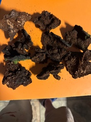
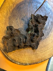
Shape and size. Irregular, lumpy, sometimes elongated along the trunk, sometimes a rounded knob. Anything from 5 cm to half a metre across. There is no regular outline and no bilateral symmetry.
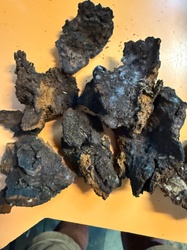
Outer crust. Coal-black to very dark brown, hard, and split into angular blocks and deep fissures like dried mud or charred timber. The crust is thin — usually only a few millimetres — and brittle, and it flakes when struck.
Attachment. It erupts through the bark rather than sitting on it, and the bark is pushed up and torn around the margin. There is no stalk and no clean line of attachment.
Interior. Break a piece off and look at the fresh face. Beneath that black crust the tissue is a striking rusty orange-brown to golden reddish-brown, corky to slightly punky, and marbled with paler cream to yellowish veins running through it. That colour contrast — black outside, rust inside — is the single most reliable field character of the species.
Where Are the Pores?
There are none, and their absence is part of the identification. Turn the conk over as best you can and check every face: a genuine Inonotus obliquus conk has no tube layer, no pore surface, and no smooth hymenium anywhere on it. Any structure with a defined layer of pores on its lower side is a bracket fungus of some other kind. Because the conk is sterile, it also never releases spores, which has consequences for the spore print discussed below.
The Fertile Stage Almost Nobody Sees
The true fruiting body appears only after the host dies or the affected part of the trunk is killed. It forms as a resupinate crust — a flat sheet — developing between the wood and the bark, which it then pushes off in plates so that the sheet is exposed. Fresh material is pale brown to greyish-brown or ochre, with a fertile surface of small angular pores, roughly three to five per millimetre, sitting over a tube layer a few millimetres deep. The tubes are characteristically set at an oblique angle to the surface, which is where the epithet obliquus comes from, and the sheet often has a paler sterile rim. Microscopically the hymenium carries hooked setae, and the spores are broadly ellipsoid, thin-walled, and pale yellowish, about 8 to 10 by 5 to 7 micrometres, browning slightly in mass. This stage decays and is broken down by insects within weeks, so encountering it is a matter of luck and timing.
Interior, Texture and Colour Change
Cut into the conk with a sturdy blade. The tissue is hard, corky and fibrous, and it resists a knife far more than any fleshy fungus. There is no latex, no gel and no rapid colour change on cutting; the rust-brown interior simply darkens gradually as it dries. Old conks become progressively harder and more deeply fissured. The mass is dense and heavy for its size, and pieces broken off leave a granular, crumbly surface.
Spore Prints and Why This One Is Different
You cannot take a spore print from a Chaga conk. This is not a limitation of technique — the conk is sterile tissue and produces no spores at all. If you set a piece on paper under a bowl overnight, you get nothing but a few dust particles, and that null result is a genuine confirmation rather than a failure. In practice it separates the conk from every poroid bracket that might be mistaken for it, all of which will drop a visible deposit given a few hours in a humid chamber.
If you find the resupinate fertile stage on a dead birch, a print is possible: cut out a section of the poroid sheet, hold it pore-side down over white paper, cover it, and leave it overnight. The deposit is pale yellowish to pale brown.
Substrate, Host Range and Season
Inonotus obliquus is overwhelmingly a fungus of birch. In the boreal and cool-temperate north it colonises living Betula through wounds and rots the heartwood over decades, the conk enlarging year after year while the tree slowly declines. It occurs less commonly on alder, beech, hornbeam and occasionally elm. Because the conk is perennial and non-seasonal, it can be found in any month, including deep winter when the bare canopy makes trunk-scanning easiest. The fertile stage, in contrast, is strictly seasonal and appears on dead or dying stems, most often in late spring and summer.
Separating It From Look-Alikes on Morphology Alone
Burls and galls. The commonest confusion. A burl is a swelling of the tree itself: it is covered in more or less continuous bark, it is wood-hard right through, and cutting into it shows swirled woody grain, never a rust-brown corky interior. No black cracked crust.
Fomes fomentarius. Very common on the same birches, but it is a hoof-shaped bracket with a smooth grey banded upper surface and a flat, pale brown pore surface underneath. Symmetry plus pores rules it out.
Phellinus igniarius. The closest genuine mimic, since it is hard, blackish and cracked with a rusty-brown interior. The difference is form: Phellinus makes a distinctly hoof-shaped or shelving bracket with a well-defined pore layer along its underside, whereas the Chaga conk is a shapeless mass with no pores anywhere.
Daldinia concentrica. Rounded, smooth, hemispherical black lumps, usually on ash. Cut one and the interior shows concentric silvery-black growth rings, not orange fibrous tissue.
Kretzschmaria deusta. A thin, brittle, black crust hugging the base of beeches and other hardwoods at ground level, grey and pale when young. Flat and crust-like rather than a bulging mass, and never up the trunk.
Black knot, Apiosporina morbosa. A black, cracked swelling, but it encircles and elongates along small twigs and branches of cherries and plums, is much smaller, and is olive-green and velvety when young.
Fire scars and charred bark. Flat, following the bark surface, with normal wood beneath.
A Field Sequence That Works
- Note the host — birch first, and a living or recently dead trunk.
- Confirm a shapeless, non-symmetrical mass erupting through torn bark.
- Confirm the crust is black and deeply block-cracked.
- Break a corner: the interior must be rusty orange-brown with cream veining.
- Check every face for pores. There must be none.
- Confirm the null spore print if there is any doubt.
Common questions
How do I tell a Chaga conk from a birch burl?
Why does a Chaga conk never produce a spore print?
Which trees carry Inonotus obliquus?
Is there a season for finding it?
Full reference entry for Chaga
Cap and fruiting body, spore print colour, substrate, season, ecological role and the species most often confused with it.
Open the Chaga entry →