Identification guide
Mica Cap Identification Guide
A tawny, deeply pleated little inkcap that fruits in dense troops around hardwood stumps, dusted when young with glistening mica-like veil granules. This guide covers the cap, the deliquescing gills and the very close relatives that share its habit.
First Impression and Setting
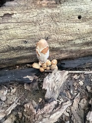
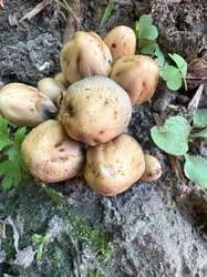
Mica cap is a mushroom of dead hardwood, and the first thing you register is the sheer number of them. Around the base of a stump, along a fallen trunk, in a wood-chip bed, or apparently straight out of a lawn where an old tree once stood, dozens to hundreds of small tan bells crowd together in a dense troop. Individual fruit bodies are only a few centimetres tall, but the colony can cover a square metre.
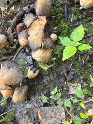
In early morning light the young caps look as if they have been dusted with ground glass. That glitter is the second immediate clue and gives the species its name. Come back in the afternoon of the same day and many of the caps will have flattened, blackened at the margin, and begun to dissolve, because these are short-lived mushrooms that complete their cycle in about a day.
The combination of clustered growth on or near hardwood, small tawny deeply grooved caps, and a glittering young surface is close to diagnostic in the field. The remaining work is separating it from two or three very close relatives, which is discussed below.
Reading the Cap
The cap is 1 to 4 cm across when expanded, occasionally 5 cm. Young caps are ovoid or egg-shaped and taller than wide, sitting like small acorns on their stems; they then open through bell-shaped to broadly conical, and finally flatten with the margin turning upward and splitting.
Colour is warm tawny to honey brown or ochre, distinctly darker at the centre, paler and often almost buff toward the margin, and fading as the cap expands and dries. Old caps go greyish and finally blacken from the edge inward as the gills liquefy.
The surface is deeply and finely grooved. Radial lines run from the margin most of the way to the disc, corresponding to the gills beneath, so the cap reads as pleated rather than smooth. Only the extreme centre stays uncreased.
Over that pleated surface, young fruit bodies carry the mica granules: minute, glistening, colourless to pale ochre particles, remnants of the universal veil. Under a hand lens they look like scattered flakes of sugar or crushed glass. They are shed easily, so rain, handling or simple ageing removes them, and a specimen may be completely smooth by midday. If your material has lost them, look for the youngest, most tightly closed caps in the colony, or check for a few granules still adhering in the grooves and near the disc.
The Gills
The gills are crowded, narrow and quite thin, attached narrowly to the stipe or nearly free. They run through a clear colour sequence: white in the youngest caps, then pale grey, then dark grey-brown, and finally black as the spores mature.
The defining behaviour is deliquescence. As the gills mature they autodigest, breaking down from the cap margin inward into a dark inky liquid that drips from the edge and stains whatever it lands on. This species deliquesces less thoroughly and more slowly than some of its relatives, so it is common to find caps that have flattened, blackened and shrivelled without turning entirely to fluid. A colony of mixed ages showing white, grey and black gills side by side is a very characteristic sight.
Because the gills disintegrate, examine young or midway specimens rather than collapsed ones when assessing gill colour and attachment.
Stipe, Base and Flesh
The stipe is 3 to 8 cm long and only 2 to 5 mm thick, straight or slightly curved, white, hollow, and very fragile. The surface is smooth to finely silky-fibrillose, sometimes with fine white down when young, and it snaps rather than bends.
There is no ring and no volva. At the very base there is often a small white mycelial pad or a few fine strands binding the stipe to wood, wood chips or soil. Trace that attachment down: even when the mushrooms appear to come out of a lawn, careful excavation usually finds buried roots or an old stump beneath.
Check for one further feature at the base of the colony: some close relatives grow from a conspicuous orange-brown to rusty felted mycelial mat called an ozonium, spread over the wood or nearby substrate. Its presence or absence is a useful character, and it is easily missed if you pick individual fruit bodies without looking at the substrate.
The flesh is thin, whitish and insubstantial. It shows no colour change on cutting apart from the general blackening of maturity, and there is no latex and no distinctive odour.
Taking and Reading a Spore Print
Timing matters more than technique here. Choose a cap that has opened but has not begun to blacken at the margin, cut the stipe off flush, place the cap gills-down on white paper, cover it with a bowl and check after two to three hours. Leave it too long and the gills liquefy onto the paper, giving a smeared black stain rather than a print. Setting up three or four caps of different ages at once is the reliable approach.
The print is very dark: blackish brown to black. Under the microscope the spores are smooth, dark reddish brown in water, roughly 7 to 10 by 4.5 to 6 micrometres, and equipped with a central germ pore. Spore shape is the practical key to separating this species from its nearest relative, being distinctly mitriform, that is, shaped rather like a bishop mitre, with a slight angular profile rather than a plain ellipse.
Substrate, Season and Ecology
This is a wood-decay saprotroph specialising in dead hardwood: stumps, fallen trunks, buried roots, wood-chip mulch, and the ground immediately around all of these. It is common in gardens, parks, hedgerows and urban plantings as well as in woodland, and it is one of the most frequently encountered fungi in landscaped mulch beds.
The season is long. Fruiting can occur from spring through late autumn and, in mild climates, in mild spells in winter, with flushes appearing within a few days of rain. The same stump will produce repeatedly across many seasons until the wood is exhausted.
Because each fruit body lasts around a day, the visible colony changes character hour by hour, and revisiting a stump over consecutive mornings is the best way to see the full developmental sequence from glittering ovoid buttons to black dripping remnants.
Separating the Closest Look-alikes
Coprinellus truncorum. The hardest separation and effectively identical in the field: same size, same tawny pleated cap, same clustered habit on hardwood, same veil granules. The reliable difference is microscopic. Its spores are ellipsoid and rounded rather than mitriform, and it typically lacks the slightly angular outline. Many field collections cannot be safely separated without checking spores, and honest recording of an aggregate is preferable to a guess.
Coprinellus domesticus and Coprinellus radians. Both are larger, with coarser and more scale-like veil remnants rather than fine mica granules, and both characteristically arise from a conspicuous orange to rusty-brown felted ozonium mat on the wood. Always look at the substrate surface around the cluster before deciding.
Coprinopsis atramentaria. Distinctly bigger and greyer, with a smooth to finely fibrillose grey-brown cap that lacks mica granules, only shallow grooving, and a more thoroughly liquefying gill mass. It tends to form clumps of a few large fruit bodies rather than dense troops of small ones.
Psathyrella candolleana. Grows in similar clustered fashion on and around hardwood stumps, but the cap is pale honey to nearly white, smooth or with only fine veil fragments hanging at the margin, and it does not deliquesce. Its gills pass from pale grey to purple-brown, and the spore print is purple-brown rather than black.
Parasola species. Very deeply pleated, membranous, almost translucent caps on thread-like stems, but they grow in grassland, lawns and dung-enriched soil rather than clustered on wood, they have no veil granules at all, and the gill collapse is minimal.
Tubaria furfuracea. Similar size and habitat on wood chips, but the gills are cinnamon brown and never blacken or dissolve, and the spore print is ochre-brown.
Coprinus comatus. No real confusion at size, but worth noting the contrast: much larger, cylindrical, shaggy white and growing from soil in grassy places.
Field Checklist
- Dense clustered troops on or around hardwood stumps, buried roots or wood chips
- Cap 1 to 4 cm, ovoid then bell-shaped, tawny to honey brown with a darker centre
- Deep radial grooving almost to the disc
- Glistening mica-like veil granules on young caps, easily washed or rubbed off
- Gills crowded, white to grey to black, partially deliquescing from the margin
- Stipe white, thin, hollow, fragile, no ring and no volva
- No orange ozonium mat on the substrate
- Blackish brown to black spore print; spores mitriform with a central germ pore
Common questions
What are the glistening particles on the cap?
The mushrooms seem to be growing in a lawn, not on wood. Is that consistent?
How is it distinguished from Coprinellus truncorum?
Why do the caps dissolve?
Full reference entry for Mica Cap
Cap and fruiting body, spore print colour, substrate, season, ecological role and the species most often confused with it.
Open the Mica Cap entry →